Research Article | | Peer-Reviewed

Surface Modification of Ti-6Al-4V Alloy by Composite Coating of Polycaprolactone Nanofibers-Fluoroapatite Nanoparticles Doped with Silicon and Magnesium

Received: 22 July 2024     Accepted: 24 August 2024     Published: 11 September 2024
Views:       Downloads:
Abstract

The purpose of this research was to create polycaprolactone nanocomposite coating - fluor apatite nanoparticles doped with silicon and magnesium, as well as polycaprolactone coating on the alloy in order to improve and modify the biological properties of this alloy. For this purpose, nano composite coating and polycaprolactone coating were first created by immersion methode. Then the physical, corrosion and biological properties of the coating created by different methods were investigated. The results indicated the creation of a uniform nanocomposite coating with a thickness of about 6.26 micrometers, with appropriate structure and phases, and an increase in roughness by adding nanoparticles to the polycaprolactone coating. Electrochemical measurements Ti6Al4V showed that the sample coated with polycaprolactone with nanoparticles has polarization RP=5.349×10⁵ Ωcm2 resistance, which is higher than the sample coated with caprolactone with polarization resistance RP=1.191×10⁵ Ωcm2 and the sample without coating with polarization resistance RP=5.2453×10⁴ Ωcm2. Cytotoxicity test showed the non-cytotoxicity of the coatings. Also, the cell growth and proliferation of the sample with nano composite coating compared to the sample without coating has a statistically significant difference. Cell adhesion on the sample with nanocomposite coating was also much better than the sample without coating and the sample with polycaprolactone coating.

Published in Composite Materials (Volume 8, Issue 2)
DOI 10.11648/j.cm.20240802.11
Page(s) 22-29
Creative Commons

This is an Open Access article, distributed under the terms of the Creative Commons Attribution 4.0 International License (http://creativecommons.org/licenses/by/4.0/), which permits unrestricted use, distribution and reproduction in any medium or format, provided the original work is properly cited.

Copyright

Copyright © The Author(s), 2024. Published by Science Publishing Group

Keywords

Surface Modification, Composite Coating, Polycaprolactone, Fluorapatite Apatite, Mtt Assay

1. Introduction
Titanium and its alloys are used for the preparation of artificial knee joints, internal fracture devices for the treatment of bone fractures, and dental implants. The tendency to use titanium and its alloys has increased because compared to stainless steel and cobalt-based alloys, they have higher corrosion resistance, higher strength-to-weight ratio, lower elasticity coefficient, and better biocompatibility. Titanium and its alloys have the highest biocompatibility among metallic biomaterials and are widely used as body implants in orthopedics and dentistry . The corrosion resistance and biocompatibility of titanium at room temperature, in the mouth and in the body of a living organism is due to the formation of a stable oxide layer with a thickness of less than one nanometer. The negative reaction of the tissue in contact with titanium or its alloys is extremely small, and bone growth or fusion with bone can occur . The chemical composition is the main and fundamental factor determining the properties and biocompatibility . For this reason, among many different types of metals and alloys, a limited number such as titanium and its alloys are known as metallic biomaterials . The chemical composition and microstructure of metal implants is the main factor determining the properties and behavior of metal in the environment, but surface conditions, cleanliness or contamination, roughness and in general surface morphology, history and how to prepare the surface and surface structure are effective in achieving the final result . The main problem of titanium and its alloys is bacterial infections and poor structural and functional connections between living bone and the implant surface . Surface preparation by creating a protective oxide layer on titanium, chemical surface treatment to increase the bioactivity of titanium and its alloys, using the sol-gel method to provide a uniform layer of titanium oxide on the surface and create a bioactive surface on titanium alloy With the method of immersion in chemical solution, the methods used to modify the surface and improve the strength of the bone graft with titanium implants are through chemical treatment and heat treatment of this implantable alloy. A review of numerous published articles and materials indicates that hydroxyapatite coatings are mainly applied on titanium or titanium alloys . Most of the attention, expectations and demands of different researchers have included bone grafting, promoting bone growth, better stability and stabilization of implants in the body, and creating an adhesive and biological interface . Increasing the corrosion resistance, reducing the corrosion current density in the body has been the attention and opinion of different researchers . Substitution of different ions in the structure of hydroxyapatite in order to provide new desirable properties or to optimize some existing properties has attracted the attention of many researchers in recent years. Based on the results of the research, replacing the hydroxyl group by fluorine ion increases the crystallinity and improves the chemical stability of the compound in the acidic environment, as well as reducing its dissolution rate in the physiological environment of the body and increasing the thermal stability . Also, the replacement of magnesium and silicon instead of calcium and phosphate ions in the structure of fluor apatite increases the solubility and absorption of calcium ions on the surface of the FA-Si-Mg material . Polymer coatings are also used on metal implants. There are several reasons, including creating anti-wear properties (using polyethylene coating with ultra-high molecular weight on titanium), creating corrosion resistance (gelatin nanocomposite coating - fluor apatite nanoparticles doped by silicon and magnesium on magnesium alloy) . There are improvements in biocompatibility, improvement in surface chemistry, and changes in surface electrical properties for the use of these coatings on metal implants . Among polymeric biomaterials, polycaprolactone has desirable properties and a long history. The good dissolution of poly-caprolactone, low melting point and the ability to mix with different types of polymers have led to the development of the use of this polymer in medical applications. This polymer is biocompatible. The results show that this polymer has a lower degradation rate than other polymer biomaterials such as polyglycolic and polylactic acid. Among the superior advantages of this polymer compared to other biopolymers are changeable degradation kinetics, mechanical properties, ease of forming, which creates a structure with pores of the right size for cell growth and can be released in a controlled manner. The medicine is effective. It is also possible to improve the hydrophilicity, cell adhesion, or biocompatibility of this polymer, which is needed for cellular responses, by using functional groups .
One of the coating methods for plants is immersion coating, which is basically based on the formation of a solution of the coated items, and the coated surface is coated by dipping into this solution and immersing it. First, the sample is immersed in the solution. Then, when the surface of the sample is wet, a layer of the coating of the desired solution sits on its surface, and then by pulling the sample out of the coated solution, the solvents and solution additives are removed from the surface of the sample by evaporating and falling . Considering the need of titanium alloy for surface modification and also considering the unique biological properties of polycaprolactone and silicon-magnesium-fluoroapatite nanoparticles, the aim of this research is the biological-surface modification of this alloy using polycaprolactone nanocomposite coating . Fluorine apatite nanoparticles doped with silicon and magnesium and investigating its corrosion and biological properties.
2. Materials and Method
In this research, the selected composite powder containing polygaprolactone and fluor apatite nanoparticles, the method of preparation and the characteristics of each of which was explained in the previous sections, was added to the specified and optimal amount with the amount of 0.2 ml of ethanol from Merck, Germany. It was mixed and placed on a magnetic stirrer. In a solution of 2% fluor apatite, 0.3 g of polycaprolactone, 0.04 g of fluor apatite, 1.8 ml of chloroform and 0.2 ml of ethanol were used. Also, the voltage is 20 volts and the distance is 17 cm.
In a 5% fluor apatite solution, 0.3 g of polycaprolactone, 0.1 g of fluor apatite, 1.8 ml of chloroform and 0.2 ml of ethanol were tested with the same specifications as before.
In 7% fluor apatite solution, 0.3 g of polycaprolactone, 0.14 g of fluor apatite, 1.8 ml of chloroform, 0.2 ml of ethanol were tested with the same specifications as before. In order to make fluorine apatite nanoparticles doped, with Sio, caf2, a mixture of silicon titanium was poured into a planetary ball asia with a zirconia chamber with a volume of 125 ml along with four subconical balls.
3. Characterization
In order to investigate the surface morphology and homogeneity of the polycaprolactone nanocomposite coating - fluorine apatite nanoparticles doped with silicon and magnesium, as well as to determine the thickness of the coating, a scanning electron microscope (Mira 3-xum) was used. In order to check the adhesion of the coating on the sublayer of titanium alloy, the adhesion test was used according to the standard (ASTM-D3359) method. A grid with an equal number of squares was applied on the surface of the polycaprolactone-fluoroapatite nanocomposite nanocomposite coating, and then a pressure-sensitive adhesive tape was applied on the scratches and quickly removed from it, the degree of separation of the coating from the surface was The percentage is stated, so the sample that has a percentage of the coating peeling off is the best sample. The surface topography and surface roughness calculation of the tested samples were checked using an atomic force microscope (AFM, DME Dual Scope 3026).
X-ray diffraction (XRD-Bruker D&A Advance) device was used to detect and determine the phases in the coating. In this technique, a lamp Cuka with a wavelength of 1.5 angstroms and a step length of 0.02 degrees and the time of each step equal to 1 second were used. The obtained patterns were compared with the standard JCPDS diffraction patterns available in the software Expert High score. Infrared spectrometry test (FTIR, Perkin Elmer Spectum 65) with Fourier transform was used to check the functional groups in the coating. In order to evaluate and compare the corrosion behavior of the samples in the simulated environment of the body, TOEFL polarization test was used. For this purpose, a computer-controlled galvanostat / potentiostat device, equipped with corrosion software, was used. Tuberculosis included the sample as a working electrode, a platinum electrode as an auxiliary electrode, and a reference electrode. The contact surface of the samples was considered to be one square centimeter and the other surfaces of the samples were covered by a non-solvent insulating material. It should be noted that before performing the electrochemical corrosion test, only the uncoated titanium alloy sample was polished to 2000 grit. Each sample was immersed in 500 ml of simulated body fluid solution at a temperature of 37 degrees Celsius. In all cases, the electrochemical test data was recorded after one hour of immersion of the samples in the electrolyte. Then, the polarization curves were obtained in the range of 250 mV with respect to the open circuit potential with the 1 mVs scanning rate. Evaluation of cytotoxicity was performed on the cells MG63 during two days.
For this purpose, the samples were first sterilized in 70% ethanol solution for 2 hours and then in ultraviolet light for 4 hours. Then the samples were placed in a 24-well cell culture plate with complete culture medium for 24 hours. Finally, the cytotoxicity test and microscopic observations were performed. In order to grow and multiply the cells to reach the required number, they were cultured inside special flasks in the laboratory environment.
Secretion of cells /Passage of cells /Cell count /Cell culture.
Cytotoxicity test was performed on days 1 and 3 as follows Preparation of cytotoxic powder solution in sterile PBS. Removing the culture medium from the surface of cultured cells under different samples and controlling and exposing them to 500 microliters of MTT solution.
Placing for 4 hours in an incubator Co2 at a temperature of 37 degrees Celsius in order to form formazan. Removal of MTT solution and dissolution of formazan using sterile DMSO solution.
Removal of MTT solution and dissolution of formazan using DMSO solution at 490 nm wavelength by ELISA reader device. Calculate the percentage of cell viability.
4. Result
The scanning electron microscope image of the surface and cross-section of the polycaprolactone nanocomposite coating - fluorine apatite nanoparticles doped with silicon and magnesium after three immersion operations were shown in the figures below, respectively. The white particles in Figure 1 are fluor apatite nanoparticles doped with silicon and magnesium, which are placed in the polycaprolactone field. As it is known, in addition to the coating being dense and free of cracks, these nanoparticles were distributed uniformly and to some extent maintaining their size in nanometer dimensions in the coating substrate. As shown in the figure, this image has a uniform thickness of 6 micrometers. Coating density, thickness uniformity, proper secondary phase distribution, and absence of microcracks are the properties of this coating .
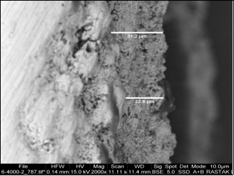
Figure 1. Electron microscope image of a) top view b) cross section of polycaprolactone nanocomposite coating - fluor apatite nanoparticles doped with silicon and magnesium.
Figure 2 shows EDAX images of uncoated, polycaprolactone-coated and polycaprolactone-nanoparticle coated samples, respectively. Numerical results obtained from these images are given in Joule 2.
After coating the titanium alloy with polycaprolactone, the surface roughness has been reduced due to the covering of grooves and surface roughness, and as a result, the surface has been smoothed. After the addition of nanoparticles to the gelatin coating, the surface roughness has increased. This increase is very small due to the small size of nanoparticles and has changed from 62 to 105 nanometers. A detailed examination of these images also shows the distribution of secondary phases and surface unevenness, which is the same as the distribution of nanoparticles in the nanocomposite coating. Viewing these images showed that the nanoparticles distributed in the gelatin bed have a good distribution.
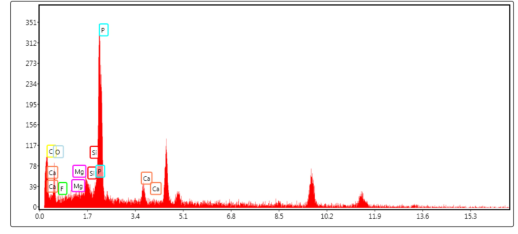
Figure 2. Energy dispersive X-ray spectroscopy images of uncoated samples / polycaprolactone coating / polycaprolactone nanocomposite coating – nanoparticles FA-Si-Mg.
Table 1. Roughness of titanium sample, polycaprolactone coating and polycaprolactone nanocomposite coating – nanoparticles FA-Si-Mg.

Sample

roughness

without any covers

321.5 nm

Polycaprolactone coating

62nm

Polycaprolactone coating - nanoparticles

105.6 nm

The next figure shows the X-ray diffraction pattern of polycaprolactone nanocomposite-fluor apatite nanoparticles doped with silicon and magnesium on titanium alloy sample by immersion method. As can be seen in this spectrum, only the peaks related to the fluorine apatite phase are observed according to the standard card. The absence of phases related to the used polymer is due to the amorphous nature of this phase and the absence of peaks related to the considerable thickness of the coating.
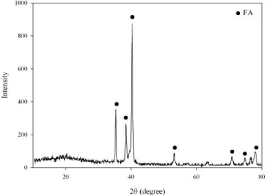
Figure 3. X-ray diffraction pattern of polycaprolactone nanocomposite coating - fluor apatite nanoparticles doped with silicon and magnesium.
The dynamic potential polarization test was started after placing the samples in the simulated solution in the body as a corrosive environment in order to reach a steady state with the electrolyte and record the open circuit potential values. The curves obtained from TOEFL polarization test are shown in the figure. The result of this test gives information about corrosion potential and corrosion current density. In the dynamic potential polarization curves of this alloy, hydrogen release and titanium dissolution occur in the range of cathodic and anodic polarization, respectively. Lower corrosion current density and higher corrosion potential means higher corrosion resistance.. As it is clear in these curves, the curves of the coated samples are inclined to the top (more positive potentials) and to the left (lower corrosion current intensity).
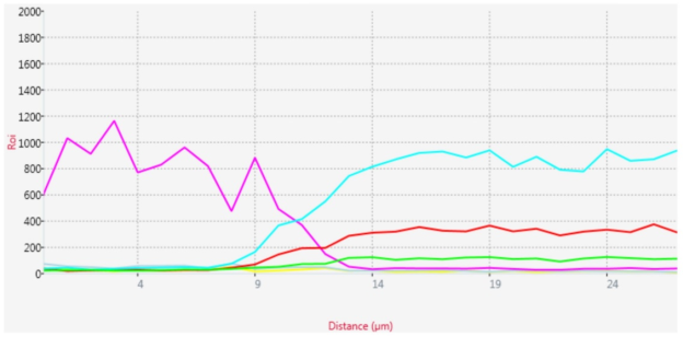
Figure 4. Corrosion test results related to samples of uncoated alloy and titanium alloy with polycaprolactone coating.
Surface chemistry and coating surface roughness are among the most important factors that control the cellular behavior of metal coatings in laboratory conditions. The normal extracellular environment contains different types of several proteins . As a result, a set of compounds with different structural, physical and chemical characteristics of visa can create ideal properties to support the growth and proliferation of cells and as a result, cell behavior is improved .
Primary cell adhesion is influenced by the surface Ti6Al4V hydrophilicity of the scaffolds, and hydrophilic surfaces create better cell adhesion than hydrophobic surfaces. The next figure shows the results of the cytotoxicity test to determine the toxicity of uncoated alloy samples, with polycaprolactone coating and the sample coated with polycaprolactone and nanoparticles after 24 hours and 72 hours of cell culture. Samples with polycaprolactone coating and nanocomposite coating do not show a statistically significant difference with the positive control, samples with polycaprolactone coating and nanocomposite coating do not show a statistically significant difference with the positive control, which means that cytotoxicity It is not visible . The growth and proliferation of cells on the sample with polycaprolactone coating has a statistically significant difference from the sample without coating, that is, the proliferation of cells on the polycaprolactone coating has increased greatly compared to the sample without coating.
Cell adhesion is an important issue because adhesion precedes other events such as cell proliferation, cell migration, and often precedes cell differentiation and function. Polycaprolactone contains many integrin binding sites for cell adhesion, migration and differentiation, which are found in polycaprolactone and other extracellular matrix proteins. Polycaprolactone contains a pseudo-sequence that improves cell adhesion and cell migration .
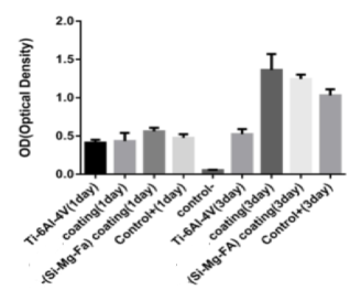
Figure 5. Cytotoxicity test results.
Figure 5 shows the scanning electron microscope images of cell adhesion on uncoated Ti6Al4V alloy, polycaprolactone-coated titanium alloy, and FA-Si-Mg nanoparticles. By creating a polycaprolactone coating on the titanium alloy, it seems that cell adhesion and cell expansion have increased due to the better cell adhesion of polycaprolactone compared to titanium. By adding silicon-magnesium fluor apatite nanoparticles, the adhesion of cells can be increased. The surface roughness and hydrophilicity of the coating increases due to the presence of nanoparticles on the surface. and improves cell adhesion of the scaffold. Better adhesion and more cell diffusion and proliferation on the surface of the nanocomposite coating can be caused by the silicon and magnesium released from the system and the improvement of the hydrophilicity of the composite composition as a result of the formation of ionic groups on the surface of the coating, which plays an essential role in promoting the differentiation of stem cells .
5. Conclusion
Polycaprolactone nanocomposite coating - fluorine apatite nanoparticles doped with silicon and magnesium by immersion method was successfully performed on titanium alloy. The nanocomposite coating with a uniform thickness with a suitable structure and chemical composition had a thickness of about 6.26 micrometers. The results of the electrochemical corrosion test showed that the titanium alloy sample with polycaprolactone nanocomposite coating - fluorine apatite nanoparticles doped with silicon and magnesium has the lowest corrosion current density among other samples. The results of the biocompatibility test in the in vitro laboratory tests showed that the titanium alloy sample coated with polycaprolactone nanocomposite - fluor apatite nanoparticles doped with silicon and magnesium showed the highest cell growth and proliferation and the best cell adhesion.
Abbreviations

PCL

Polycaprolactone

Conflicts of Interest
The authors declare no conflicts of interest.
References
[1] T. Akahovi and M. Niinomi, Fracture Characteristics of Fatigued Ti-6Al-4V ELI as an Implant Material, Materials Science and Engineering, A243(1998) 237-243, 1998.
[2] WJ. O’Brien, Dental Materials and Their Selection, 2th ed., Quintessence Publishing Co, Inc., 1997.
[3] Reneker D., Kataphinana W., Theron A., Zussman E., nanofiber garlands of polycaprolactone by electrospinning, Polymer, 43, 6785-6794, 2002.
[4] S. G. Steinmann, Corrosion of surgical implants-in vivo and in vitro tests, in: Evaluation of Biomaterials, ed. By Gd Winter, JL leray, K deGroot, John Wiley and sons Ltd, 1980.
[5] S. G. Steinmann, Corrosion of titanium and titanium alloys for surgical implants, Titanium Science and Technoloy, Munich: Deutsche Gesellschaft Fur Metallkunde e. V, 2(1985) 1373-1379.
[6] B. Kasemo and J. Lausmaa, Biomaterial and implant surfaces: on the role of cleanliness, contamination, and preparation procedures, Journal of Biomedical Materials Research, 22(1988) 145-158, 1988.
[7] J. R. Smith and D. A. Lamprou, Polymer coatings for biomedical applications-a review, Transactions of the IMF, 92(2014) 9-19.
[8] M. Shirkhanzadeh, Electrochemical preparation of protective oxide coatings on titanium surgical alloys, Journal of Materials Science: Materials in Medicine, 5(1992) 322-325.
[9] H. M. Kim, F. Miyaji, T. Kokubo, and T. Nakamura, Preparation of bioactive Ti and its alloys via simple chemical surface treatment, Journal of Biomedical Materials Research, 32(1996) 409-417.
[10] J. Breme, E. Eisenbarth, H. Hildebrand, A. Ralison, W. Schulte and J. Meyle, Modification of the surface of titanium implants for an improved osseointegration, proceeding of titanium 95, Birmingham, Uk, 2(1995) 1792-1799.
[11] H. B. Wen, J. R. de Wijn, F. Z. Cui, and K. de Groot, Preparation of bioactive Ti6Al4V surfaces by a simple method, biomaterials, Biomaterials, 19(1998) 215-221.
[12] J. M. Gomez-Vega, E. saiz,, A. P. Tomsia, G. W. Marshall, and S. J. Marshal, Bioactive Glass coatings with hydroxyapatite and bioglass particles on Ti-based implants. 1. processing, Biomaterials, 21(2000) 105-111.
[13] W. Suchanek, M. Yoshimura, Processing and properties of hydroxyaptite-based biomaterials for use as hard tissue replacement implants, Journal of Materials Research, 13(1998) 94-117.
[14] L. B. Lum, R. beirne and D. A. Curtis, Histologic evaluation of hydroxyapatite-coated versus uncoated titanium blade implants in delayed and immediately loaded applications, The International Journal of Oral & Maxillofacial Implants, 6(1991) 456-462.
[15] A. Pichat, L. M. Rabbe, J. Rieu, A. Romabert, C. Chabrol, and M. Robelet, Effect of ion implantation on titanium alloy/polyethylene and 16L stainless steel/polyethylene friction couples running in joint prosthese, Surface and Coatings Technology, 1(1991) 15-22.
[16] H. W. Kim, H. E. Kim, and J. C. Knowles, Fluor-hydroxyapatite sol-gel coating on titanium substrate for hard tissue implants, Biomaterials, 25(2004) 3351-3358.
[17] Woodruff, M. A., and Hutmacher, W. H., “The return of a forgotten polymer : Polycaprolactone in the 21st century”, Progress in Polymer Science, Vol. 35, pp. 1217–1256, 2010.
[18] A. Jafarzadeh, T. Ahmadi, M. Taghian Dehaghani, K. Mohemi, Synthesis, Corrosion and Bioactivity Evaluation of Gelatin/Silicon and Magnesium Co-Doped Fluorapatite Nanocomposite Coating Applied on AZ31 Mg Alloy, Russian Journal of Non-Ferrous Metals, 59(2018) 458-464.
[19] S. A. Sell, P. S. Wolfe, K. Garg, et al., The Use of Natural Polymers in Tissue Engineering: A Focus on Electrospun Extracellular Matrix Analogues, Polymers, 2(2010) 522-553.
[20] Z. X. Meng, Y. S. Wang, C. Ma,, et al., Electrospinning of PLGA/gelatin randomly-oriented and aligned nanofibers as potential scaffold in tissue engineering, Materials Science and Engineering: C., 30(2010) 1204-1210.
[21] H. Hajiali, S. shahgasempour, M. R. Naimi-Jamal, et al., Electrospun PGA/gelatin nanofibrous scaffolds and their potential application in vascular tissue engineering, International Journal of Nanomedicine, 6(2011) 2133-2141.
[22] 22: Detta N. et al., Melt Electrospinning of Polycaprolactone and Its Blends with Poly (ethylene glycol), Polym. Int., 59, 1558-1562, 2010.
[23] J. Degner, F. Singer, L. Cordero, A. R. Boccaccini, S. Virtanen, Electrochemical investigations of magnesium in DMEM with biodegradable polycaprolactone coating as corrosion barrier, Applied Surface Science, 282(2013) 264-270.
[24] C. Wu, Z. Wen, C. Dai, Y. Lu, F. Yang, Fabrication of calcium phosphate/chitosan coatings on AZ91D magnesium alloy with a novel method, Surface and Coatings Technology, 204(2010) 3336–3347.
[25] P. V. Kozlov, G. I. Burdygina, The structure and properties of solid gelatin and the principles of their modification, Polymer, 24(1983) 651-666.
[26] M. Djabourov, J. Leblond and P. Papon, Gelation of aqueous gelatin solutions. I. Structural investigation, Journal of Physics France, 49(1988) 319-332.
[27] M. Kazemzadeh Narbat, F. Orang, M. Solati Hashtjin and A. Goudarzi. Fabrication of Porous Hydroxyapatite-Gelatin Composite Scaffolds for Bone Tissue Engineering, Iranian Biomedical Journal 10(4): (October 2006) 215-223.
[28] X. Cui, Y. Li, Q. Li, et al., Influence of phytic acid concentration on performance of phytic acid conversion coatings on the AZ91D magnesium alloy, Materials Chemistry and Physics, 111(2008) 503–507.
[29] H. Ashassi-Sorkhabi, and M. Eshaghi, Corrosion resistance enhancement of electroless Ni–P coating by incorporation of ultrasonically dispersed diamond nanoparticles, Corrosion science, 77(2013) 185–193.
[30] Lyons J. M., Melt-Electrospinning of Thermoplastic Polymers: An Experimental and Theoretical Analysis, Ph.D. Thesis, Drexel University, October 2004.
[31] K. M. Woo, V. J. Chen, and P. X. Ma, Nano-fibrous scaffolding architecture selectively enhances protein adsorption contributing to cell attachment, Journal of Biomedical Materials Research A, 167(2003) 531-437.
[32] I. K. Kwon, S. Kidoaki, and T. Matsuda, Electrospun nano- to microfiber fabrics made of biodegradable copolyesters: structural characteristics, mechanical properties and cell adhesion potential, Biomaterials, 26(2005) 3929-3939.
[33] Y. Huang, S. Onyeri, M. Siewe, A. Moshfeghian, and S. V. M. Ã, In vitro characterization of chitosan – gelatin scaffolds for tissue engineering, Biomaterials, 26(2005) 7616-7627.
[34] N. Johari, M. H. Fathi, M. A. Golozar, Fabrication, characterization and evaluation of the mechanical properties of poly (e-caprolactone)/nano-fluoridated hydroxyapatite scaffold for bone tissue engineering, Composites: Part B, 43(2012) 1671-1675.
[35] M. Diba, M. H. Fathi, and M. Kharaziha, Novel forsterite/polycaprolactone nanocomposite scaffold for tissue engineering applications, Materials letters, 65(2011) 1931-1934.
[36] M. Kharaziha, M. H. Fathi, and H. Edris, Effects of Surface Modification on the Mechanical and Structural Properties of Nanofibrous Poly (ε-aprolactone)/Forsterite Scaffold for Tissue Engineering Applications, Material Science and Engineering C, 33(2013) 4512-4519.
Cite This Article
  • APA Style

    Amjad, N. E., Sadeghi, M., Mirbagheri, M., Vahdat, K. (2024). Surface Modification of Ti-6Al-4V Alloy by Composite Coating of Polycaprolactone Nanofibers-Fluoroapatite Nanoparticles Doped with Silicon and Magnesium. Composite Materials, 8(2), 22-29. https://doi.org/10.11648/j.cm.20240802.11

    Copy | Download

    ACS Style

    Amjad, N. E.; Sadeghi, M.; Mirbagheri, M.; Vahdat, K. Surface Modification of Ti-6Al-4V Alloy by Composite Coating of Polycaprolactone Nanofibers-Fluoroapatite Nanoparticles Doped with Silicon and Magnesium. Compos. Mater. 2024, 8(2), 22-29. doi: 10.11648/j.cm.20240802.11

    Copy | Download

    AMA Style

    Amjad NE, Sadeghi M, Mirbagheri M, Vahdat K. Surface Modification of Ti-6Al-4V Alloy by Composite Coating of Polycaprolactone Nanofibers-Fluoroapatite Nanoparticles Doped with Silicon and Magnesium. Compos Mater. 2024;8(2):22-29. doi: 10.11648/j.cm.20240802.11

    Copy | Download

  • @article{10.11648/j.cm.20240802.11,
      author = {Nasim Emammi Amjad and Mahshid Sadeghi and Mahshad Mirbagheri and Khadijeh Vahdat},
      title = {Surface Modification of Ti-6Al-4V Alloy by Composite Coating of Polycaprolactone Nanofibers-Fluoroapatite Nanoparticles Doped with Silicon and Magnesium
    },
      journal = {Composite Materials},
      volume = {8},
      number = {2},
      pages = {22-29},
      doi = {10.11648/j.cm.20240802.11},
      url = {https://doi.org/10.11648/j.cm.20240802.11},
      eprint = {https://article.sciencepublishinggroup.com/pdf/10.11648.j.cm.20240802.11},
      abstract = {The purpose of this research was to create polycaprolactone nanocomposite coating - fluor apatite nanoparticles doped with silicon and magnesium, as well as polycaprolactone coating on the alloy in order to improve and modify the biological properties of this alloy. For this purpose, nano composite coating and polycaprolactone coating were first created by immersion methode. Then the physical, corrosion and biological properties of the coating created by different methods were investigated. The results indicated the creation of a uniform nanocomposite coating with a thickness of about 6.26 micrometers, with appropriate structure and phases, and an increase in roughness by adding nanoparticles to the polycaprolactone coating. Electrochemical measurements Ti6Al4V showed that the sample coated with polycaprolactone with nanoparticles has polarization RP=5.349×10⁵ Ωcm2 resistance, which is higher than the sample coated with caprolactone with polarization resistance RP=1.191×10⁵ Ωcm2 and the sample without coating with polarization resistance RP=5.2453×10⁴ Ωcm2. Cytotoxicity test showed the non-cytotoxicity of the coatings. Also, the cell growth and proliferation of the sample with nano composite coating compared to the sample without coating has a statistically significant difference. Cell adhesion on the sample with nanocomposite coating was also much better than the sample without coating and the sample with polycaprolactone coating.
    },
     year = {2024}
    }
    

    Copy | Download

  • TY  - JOUR
    T1  - Surface Modification of Ti-6Al-4V Alloy by Composite Coating of Polycaprolactone Nanofibers-Fluoroapatite Nanoparticles Doped with Silicon and Magnesium
    
    AU  - Nasim Emammi Amjad
    AU  - Mahshid Sadeghi
    AU  - Mahshad Mirbagheri
    AU  - Khadijeh Vahdat
    Y1  - 2024/09/11
    PY  - 2024
    N1  - https://doi.org/10.11648/j.cm.20240802.11
    DO  - 10.11648/j.cm.20240802.11
    T2  - Composite Materials
    JF  - Composite Materials
    JO  - Composite Materials
    SP  - 22
    EP  - 29
    PB  - Science Publishing Group
    SN  - 2994-7103
    UR  - https://doi.org/10.11648/j.cm.20240802.11
    AB  - The purpose of this research was to create polycaprolactone nanocomposite coating - fluor apatite nanoparticles doped with silicon and magnesium, as well as polycaprolactone coating on the alloy in order to improve and modify the biological properties of this alloy. For this purpose, nano composite coating and polycaprolactone coating were first created by immersion methode. Then the physical, corrosion and biological properties of the coating created by different methods were investigated. The results indicated the creation of a uniform nanocomposite coating with a thickness of about 6.26 micrometers, with appropriate structure and phases, and an increase in roughness by adding nanoparticles to the polycaprolactone coating. Electrochemical measurements Ti6Al4V showed that the sample coated with polycaprolactone with nanoparticles has polarization RP=5.349×10⁵ Ωcm2 resistance, which is higher than the sample coated with caprolactone with polarization resistance RP=1.191×10⁵ Ωcm2 and the sample without coating with polarization resistance RP=5.2453×10⁴ Ωcm2. Cytotoxicity test showed the non-cytotoxicity of the coatings. Also, the cell growth and proliferation of the sample with nano composite coating compared to the sample without coating has a statistically significant difference. Cell adhesion on the sample with nanocomposite coating was also much better than the sample without coating and the sample with polycaprolactone coating.
    
    VL  - 8
    IS  - 2
    ER  - 

    Copy | Download

Author Information